CBLO and TPLO surgery
Key Points:
- CBLO and TPLO surgery both use a radial cut osteotomy to level the tibial plateau.
- More invasive than TTA surgery however has a better outcome with dogs greater than 30kg or tibial slope greater than 25 degrees.
- Postoperatively there are weekly visits for 4 weeks. On leash exercise can generally be started at 1-2 weeks post operatively. A radiograph is taken at 8-12 weeks to ensure the osteotomy has healed at which point the dog can go to full off leash exercise.
CBLO was developed as a refinement of the concepts of TPLO and addresses some of the issues that could cause complications with TPLO particularly in certain animals. One of the issues with TPLO is that in a lot of dogs the end result is that the load bearing axis of the tibia is moved further away from the anatomical axis of the tibia. CBLO addresses this by inverting the rotation which results in the weight bearing axis being brought into alignment with the anatomical axis.
Surgery description
Your pet is anaesthetised ready for surgery and the area clipped and prepared for sterile surgery. The stifle is approached through a medial incision (inside of the leg). The subcutaneous tissues are dissected and the stifle joint is entered. The joint and menisci are inspected. Damaged menisci are removed and the joint is then closed with high grade synthetic absorbable suture material. Once the joint is inspected the osteotomy is performed. The location of the radial cut is determined from pre operative planning radiographs and the cut made. The proximal segment is then rotated a set distance determined from pre operative radiographs. The rotated segment is then fixed in place with special screws and plates. The wound is closed and post operative radiographs are taken to ensure correct placement of all the implants.
What to expect from CBLO / TPLO surgery
Most studies and our clinical experience has been that over 95% of clients regard results of surgery as “excellent”. Obviously one persons assessment of excellent is different from anothers however our impressions are that there is a more normal function and better long term relief from symptoms achieved with this surgery compared with other treatment options including conservative (i.e. non surgical) management, graft repair or DeAngelis suture repair. Whilst TTA will often have a slightly faster recovery, long term studies indicate CBLO/TPLO has better long term results with fewer complications than TTA particularly in dogs over 30kg or with very steep tibial slopes.
We CANNOT CURE ARTHRITIS!! If your pet has significant arthritic changes in the joint then results will not be as good as if surgery was done earlier. Significant degeneration of the joint can occur in as little as 4-6 weeks! This is why we recommend surgery as soon as possible after injury.
Does my pet need any care after surgery?
We will usually keep your pet in overnight after surgery for monitoring. Your pet will go home the following day and will need to take antibiotics and pain relief medications for 10 to 20 days.To get the best results from the surgery your pet needs to have follow up visits weekly for 4 weeks. At these visits they will receive an injection (Cartrophen) to help the joint recover from surgery. Additionally they will have physiotherapy and you will be given exercises to do with them at home to help with recovery.
We typically expect your pet to be 20-30% weight bearing at 1 week post op, 40-50% weight bearing at 2 weeks post op, 60-70% weight bearing at 3 weeks post op and around 80% weight bearing at a walk 4 weeks post op. If there is meniscal damage then that will typically delay recovery by 1-2 weeks. We do not recommend off leash exercise until radiographs at 8-12 weeks post op show good healing of the osteotomy.
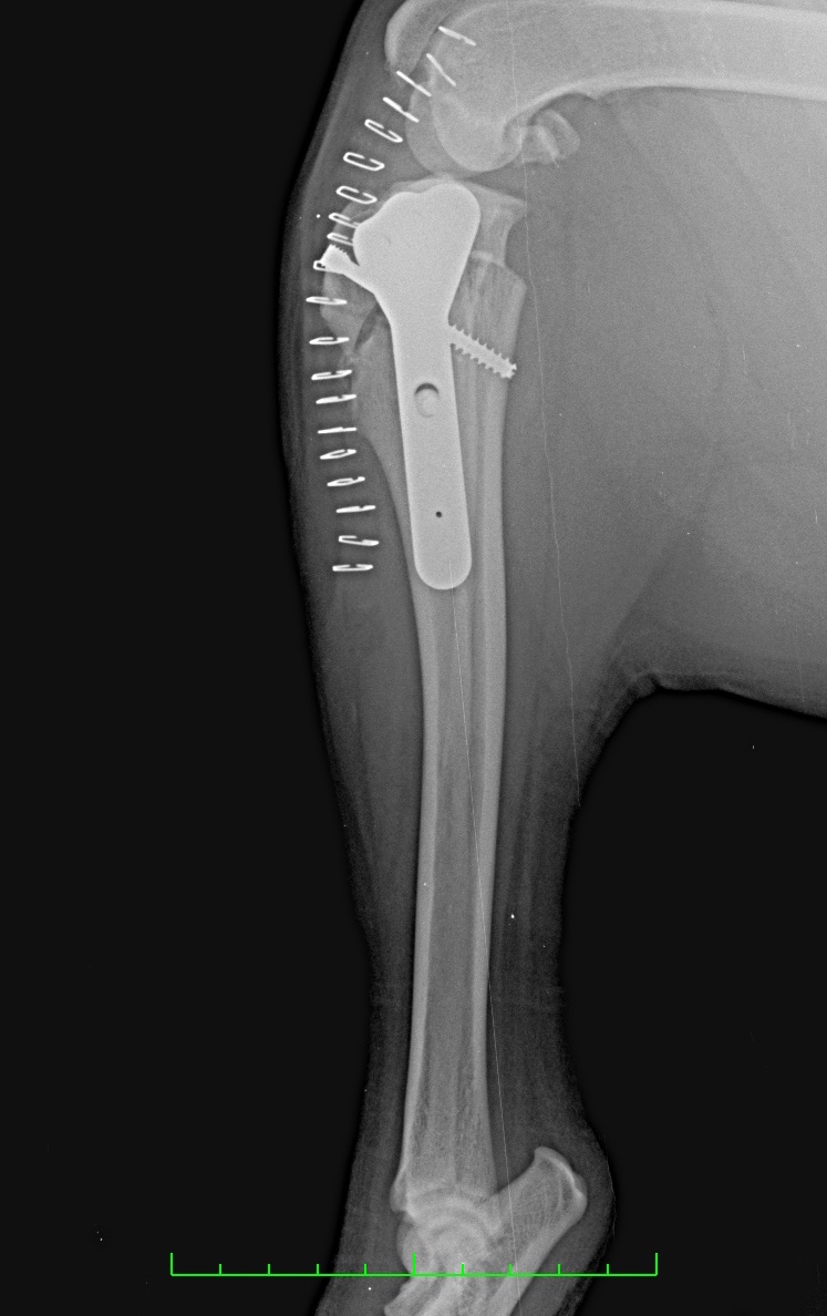
Post operative radiograph of CBLO surgery

Post operative radiographs of TPLO surgery
Cruciate Disease in Dogs
Key Points
- Cruciate disease is the most common orthopaedic injury in dogs. Over 90% of hind leg lameness in dogs is due to cruciate disease.
- The cause of cruciate disease is unclear with a number of theories, in all likelihood there are multiple causes. The disease is however a degenerative meaning it is more common in middle age to older dogs.
- Once damaged the ligament never repairs itself and requires surgery to return the stifle to good function. Modern surgical repairs can restore up to 90% of pre injury function to the leg.
- Left untreated the stifle will develop severe arthritis that causes long term pain and discomfort.
What are the Cruciate Ligaments?
The cruciate ligaments occur in the stifle (knee) of all mammals and help to stabilise the stifle. They stop the femur and tibia from moving forward and backward relative to each other and ensure that the stifle essentially acts like a hinge.
What is Cruciate disease?
Unfortunately it is very common for these ligaments to become damaged thus destabilising the stifle. Cruciate disease is easily the most common orthopaedic condition in dogs. In our practice it would account for over 70% of all lameness and arthritis problems in all breeds. Despite this fact and many years of research it is still unclear as to the exact causes of cruciate disease in dogs. In all likelihood cruciate ligament damage is a consequence of the combination of a variety of factors including:
- Steep tibial slope placing constant pressure on the Anterior / Cranial Cruciate Ligament (ACL). This is the theory underpinning most of the modern surgical treatments for ACL damage in dogs.
- Generalised degenerative joint disease associate with excess body fat tissue.
- Traumatic over extension or twisting of the stifle.
- Poor blood supply to the ACL.
Whilst we don't necessarily know the exact cause of ACL damage in the dog what we do know is that once damaged the inflammation generated causes more damage causing more inflammation in a self perpetuating cascade. We also know that the ACL once damaged essentially never heals or repairs itself to any significant degree.
How do I know if my dog has Cruciate Disease?
Well, the simple answer is if your dog is lame in a hind legs for more than a couple of days then it probably has damaged it's ACL! There are however some other problems that can cause hind leg lameness so it is important that your dog is assessed by a veterinarian.
There are a number of characteristics of dogs with cruciate disease. The stifle can be swollen, there may be swelling on the inside of the tibia (called “medial buttress”), many dogs stand or walk with the good leg swinging inwards to be directly under the body as opposed to under the hip, and many dogs will sit with their good leg tucked underneath them and the bad leg stretched out to the side (the “sit” test). Essentially though the diagnosis is made by feeling the stifle for instability and cranial drawer (or forward movement of the tibia compared to the femur). Sometimes however this movement is very subtle and if the dog is very tense it can be hard to feel this. Consequently your veterinarian may suggest sedating your dog to check the stifle and also to get radiographs of the leg.
Does my dog need X-rays?
Whilst radiographs are not essential for the diagnosis of cruciate disease in the dog they are helpful to rule out other diseases that may be occurring at the same time. Occasionally we also get very swollen stifles where it is difficult to feel a cranial drawer and the swelling could be due to growths or other problems and radiographs in these cases help to rule those other causes out and confirm a diagnosis of ACL damage. Radiographs are also important for planning the surgical repair.
Treatment
Medical
Unlike humans cruciate disease in the dog cannot be treated with medical management. It just doesn't work. While in some cases some short term pain relief can be achieved the stifle will continue to be unstable and inflamed developing significant amounts of scar tissue and fibrosis that restricts the movement of the stifle and the leg. In certain cases, very old animals where life expectancy is likely to be less than 12 months then the use of anti inflammatory medications, joint supplements and pain killers can provide reasonable quality of life for a short period of time. Long term use of medical management is ineffective, risks causing side effects and over a period of any longer than 2-3 years is likely to cost just as much as surgery.
Surgical
There is little doubt that modern surgical techniques are the best option for treating cruciate disease in dogs. Older techniques including graft repairs and De Angelis or Lateral suture techniques provided some short to medium improvement however studies have clearly demonstrated that after 12 months both those techniques have failed and the stifle continues to be unstable and the joint chronically inflamed. What are known as tibial plateau slope modifying techniques e.g. TTA, TPLO, CBLO have revolutionised treatment of cruciate disease in dogs and have meant that we can now restore up to 90% of pre injury function long term in a majority of dogs. Kate Toyer our surgeon is trained and has performed all of the modern tibial osteotomy surgeries (and most of the older surgeries as well!). There is no one particular surgery which is the best and each have their advantages and disadvantages. The principle determining factor for which surgery is best is the size of the dog and what is known as the tibial slope determined from radiographs. After assessing radiographs and your dog clinically Kate will recommend what she believes to be the best surgery for your dog.
For more details on these surgeries please see our page here.
Fracture repair surgery
- Fracture repair when performed correctly will successfully heal over 90% of fractures with no long term consequences or adverse effects even in severe multiple fragment fractures.
- Correct fracture repair requires a full assessment of the animals health overall as well as a complete assessment of the fracture both clinically and radiographically.
- There are a number of different techniques for repairing fractures including various bone plating systems, conventional, circular and hybrid external fixators, interlocking nails to name a few, each with it's advantages and disadvantages. The best technique for a particular fracture is based on clinical and radiographic assessment.
- Due to the high morbidity and long term complication rate of poor healing we no longer recommend any form of “casting” for fracture stabilisation.
- Adequate postoperative care and follow up assessments are crucial to minimise complications and get the best outcome for the animal.
Bone fractures are highly traumatic and often very painful injuries. In the past repair and healing often required long periods of forced rest or non weight bearing on the affected bone as we attempted to reconstruct the bone and literally almost glue it together in what we believe was the best way to repair fractures. Unfortunately complications were common and could sometimes lead to non functional bones and require amputation of limbs.
Thankfully our understanding of bone healing and biomechanics has improved dramatically in the last 20 years. We now know that piecing together a shattered bone is not only not effective, the handling of fragments may decrease their ability to heal. The focus of modern fracture repair is now to transfer weight bearing away from the fracture location and to maintain the orientation of joints above and below / either side of the fracture. Fragments are generally left alone for the body to heal with it's own natural processes and surgical techniques to minimise interference to blood supply are employed.
Fracture Types
Fractures can be broadly classified into:
- Open or Closed. An open fracture is where the bone has been exposed to the environment and possible infection. Any fracture where the surrounding skin has been damaged should be treated as possibly open. Open fractures are far more prone to infection and ideally implants should not be left in the animal if a possible infection is suspected.
- Simple or comminuted. This is the number of pieces the bone has fractured into. If there is only 1 fracture line with 2 pieces, or perhaps a 3rd large fragment, then the fracture is known as simple. If however there are multiple fragments, particularly small fragments, then it is known as comminuted. Simple fractures can be pieced back together with minimal complications however if the are multiple small fragments then the aim is to minimise handling of those fragments as much as possible.
Fracture repair techniques
It is generally acknowledged that successful complication free healing for fractures requires surgical intervention and the use of implants to support the fractured limb. There are however a variety of implants and implant systems, each with their advantages and disadvantages. The best option is often a combination of a variety of factors and weighing up the pro's and con's of each technique.
Bone Plates
Bone plates and screws have been the mainstay of fracture fixation for the last 40 years in both human and veterinary fractures. Recent advances in the development of locking bone plates will in all likelihood mean they continue to be the preferred method of fracture fixation for most fractures. The technique consists of securing a rigid metal plate to the bone with screws. The plate serves two functions, firstly it provides a rigid mechanism to transfer weight from one side of the fracture to the other, and secondly it brings the fragments into alignment to allow healing in an anatomically correct position.
Pros
- Single surgery definitive repair. i.e. their is usually no requirement for additional surgeries to remove implants.
- Anatomical reduction and alignment generally very accurate.
- High rigidity particularly of new Locking plate technology means that animals are often weight bearing almost immediately after surgery.
Hip Dysplasia
Key Points:
- Hip dysplasia (HD) is DJD of the hip joints caused by a combination of genetic and environmental / dietary factors.
- Specialised distraction radiography (PennHIP or DLS techniques) can predict the likelihood of HD development from the age of 6 months.
Principally a problem affecting dogs (though it can affect cats), hip dysplasia has been recognised for many years in the veterinary literature as far back as the 1950's as a collection of radiographic and clinical signs associated with reduced function and degeneration of the coxofemoral joint. Unfortunately incorrect interpretation of the original descriptions by both lay people and the veterinary community led to the term Hip Dysplasia being used as a definition of an etiologic agent rather than a description of a clinical syndrome. There is little doubt now that there are underlying abnormalities of growth and function of the coxofemoral joint that result in degeneration and reduced function of the joint. There are a number of genetic and environmental factors that contribute to this degeneration but they can be broadly broken down into the following groups:
- Factors affecting hip laxity. These are principally genetic BUT excessive weight or physical activity can contribute to stretching a weak coxofemoral joint.
- Factors affecting cartilage and joint biology. These are mostly environmental (e.g. diet) however there is some contribution from genetic factors such as poor capacity to produce proteoglycans.
The combination of these factors causes progressive damage to the cartilage of the hip joint result in arthritis, restriction of movement in the hip and pain.
Clinical signs
Clinical signs can be varied and not all dogs show all signs. The most common signs are lameness, “bunny hopping” when running, reluctance to jump, pushing up with the front legs when getting up, slow to sit down, and reluctance to exercise. Most clinical signs with hip dysplasia are progressive and fairly gradual i.e. if the dog (or cat) is suddenly lame and won't put a foot down that is unlikely to be hip dysplasia.
Diagnosis and radiography
Clinical signs of hip dysplasia can be similar to those of other orthopaedic diseases in dogs and cats as well. Notably cruciate ligament disease and to a lesser extent spinal disease can cause some similar clinical signs. If you suspect your pet has hip dysplasia then it is important to get them examined by a veterinarian and also radiographed to determine if the hips are the cause of the problem and how severe the damage is.
Unfortunately in the past radiographic techniques to assess hip dysplasia really only detected the problem after it had occurred and it was too late to intervene. Recent advanced distraction radiograph techniques such as PennHIP or DorsoLateral Subluxation (DLS) can accurately assess hip laxity and can predict the development of hip dysplasia from as early as 16 weeks and at 6 months have an almost 95% correlation with onset of clinical signs. These techniques measure how loose the hip joints are by putting pressure on the hips to try and distract them out of the joints. In addition as opposed to older techniques that were a sort of yes / no diagnosis of hip dysplasia, PennHIP and DLS actually give an indication of the severity of hip laxity and the likely age of onset of problems.
Treatment
Treatment has traditionally been based around medical management of the DJD until the animal is no longer responsive at which point the only treatment option available is Total Hip Replacement (THR). The ability to diagnose a predisposition to hip dysplasia with PennHIP or DLS has meant that we have the option to perform procedures that can actually prevent hip dysplasia from occurring. The two procedures that have been most effective are Juvenile Pubic Symphysiodesis (JPS) and Triple / Double Pelvic Osteotomy (TPO/DPO).
Medial Patella Luxation (MPL)
Key Points
- Medial Patella Luxation is an anatomical abnormality that causes the kneecap to dislocate inwards.
- MPL is a congenital developmental condition that progresses in severity with age.
- Surgical realignment of the patella is a highly effective treatment particularly when performed early in the animals life before arthritis develops from the constant wear and tear.
- Surgical correction needs to consist of either / both of Tibial Crest Translocation (TCT) and Trochlear Groove deepening / resection (TGR).
MPL is the most common orthopaedic problem in small breed (<10kg) dogs. It affects the stifle (knee) joints of the hindlegs and results in chronic intermittent luxation of the patella (knee cap). The essential anatomical abnormality is that the Tibial Crest is medially deviated, i.e. it is positioned too far towards the inside of the leg. Because the tibial crest is the point of attachment for the patella ligament this medial displacement pulls the patella medially, towards the inside of the leg as the dog walks. The patella constantly being dragged across the medial ridge of the trochlea results in the medial ridge being worn down and reducing it's resistance to the patella luxating out of the trochlea groove. Eventually the constant wear and tear results in significant arthritic changes in the stifle and pain and decreased function of the hind leg.
Clinical Signs
Some dogs may have no apparent clinical signs and even dogs with severe MPL seem to run around just fine, particularly if they are younger. That being said the most common clinical sign is “skipping” or the dog holding the leg up for 2 or 3 strides before going back to a normal gait. Usually this is intermittent and is caused by the patella locking in the dislocated position until the dog manages to manipulate the leg to get it back into a normal position. Other less frequent signs include a reluctance to jump or in severe cases a bandy legged appearance of the hind legs.
It is important to note that the disease is typically progressive i.e. a dog that has a mild MPL at a checkup will almost always get worse. This can occur quite quickly, over a period of 6 months, or it can be many years. Some dogs have low grade MPL their entire life without having clinical signs or needing treatment.
Grading
Patella luxation is graded according to an internationally recognised grading system.
- Grade 0: Normal. Patella cannot be manipulated out of the trochlea groove.
- Grade 1: IN/IN. Patella can be manipulated out of the trochlea groove but returns immediately once released.
- Grade 2: IN/OUT. Patella can be manipulated out of the trochlea groove and remains out once released. Movement or manipulation of the leg will eventually cause the patella to return to the trochlea groove.
- Grade 3: OUT/IN. Patella is out of the trochlea groove but can be pushed in to the trochlear groove. Movement or manipulation of the leg will eventually cause the patella to move out of the trochlear groove again.
- Grade 4: OUT/OUT. Patella's out of the trochlear groove and cannot be manipulated in to the trochlea groove. In many cases like this the trochlea groove is so worn that it barely exists.
Treatment
As the underlying problem is anatomical treatment involves surgical realignment of the patella mechanism. Occasionally in very old animals (>14yrs) we may suggest medical management of the arthritis rather than surgery however most animals initially present with clinical signs requiring treatment at under 7 yrs of age and early treatment maximises long term outcome.
The current recommendations of the American, European, Australian and New Zealand Colleges of Veterinary Surgery for dogs with MPL are:
- a grade 1 or 2 MPL with no clinical signs should be monitored and reassessed every 6 months.
- Dogs with a grade 2 MPL and clinical signs and ALL dogs with grade 3 and 4 MPL should have surgical realignment of the patella irrespective of whether they are showing clinical signs or not.
- Surgical realignment should consist either a TCT, TGR or both.